Life and Liberty
For Women
The Truth About Anti-abortion Pictures
The Truth About Anti-abortion Pictures of Alleged Aborted Fetuses
by Sara B.
Journalist and Volunteer, Life and Liberty for Women
Since the genesis of the anti-abortion campaign, pro-life activists have used pictures to further their cause. However, many of these visuals present glaring contradictions and inconsistencies that have long been ignored (perhaps because of the delicate nature of the debate). Life and Liberty for Women is willing to call these propagandists out on their half-truths and blatant lies.
Misrepresentation
A considerable number of anti-abortion visuals feature an almost fully developed fetus. Abortions preformed at this stage via hysterotomy or D&X abortions are rare. Only about 1.5% of abortions are performed at 21-weeks or older, according to according to a 2000 study conducted by the Nation Center for Chronic Disease (CDC). A July 1992 LIFE magazine article, The Great Divide, reported that Reverend Robert Schenk, member of anti-choice coalition Operation rescue, attended a demonstration outside an abortion clinic in Buffalo, NY, with "Baby Tia", a 7-inch, gray-tinted and formaldehyde-soaked dead fetus. In the escalating madness of the crowd, the fetus was dropped onto the sneaker-trodden street. Authorities arrested Schenk and confiscated the fetus, which was taken to a coroner, only to be identified as an approximately 20-week-old stillborn. The article reads, "Many pro-choice supporters in Buffalo are angry about the distance between their reality-what they see in the clinics-and the images the anti-abortionists present as fact. 'What they are showing to the public is a lie,' says Joni Ladowski, a nurse at a nearby clinic, as she unfolds a length of gauze. In the center lies what appears to be a clot the size of a peanut. It is a fetus, nine weeks old. 'This is an abortion,' she says."

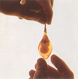
Tear-drop baby
First used in the anti-abortion campaign by Dr. J.C. Willke, president and founder of International Right to Life, in his 1971 book, Handbook on Abortion, this picture has circulated through anti-choice brochures and websites without much question of its origin or accuracy. This picture depicts a ruptured tubal pregnancy that Willke would like you to believe is 6-weeks-old.
A 6-week-old fetus, from crown-to-rump, measures 4-5 mm, according to William's Obstetrics, or 1/6-1/5 inch (the size of a BB pellet). The fetus in the photo, when compared to the fingers holding it, appears to be about an inch in length or the size of an 8- to 10-week-old fetus.
We have also posted a photo of a 6-week-old fetus that was published in A Child is Born, by Lennart Nilsson. The two are demonstrably different! Accurately labeling the fetus's gestational age is important because this photo represents the mortality of developing life. One may look at this picture and see a fetus only into its second month of development and think, "look how far it's come." But when labeled accurately, this fetus would be approaching its second trimester, a period when only 12% of abortions are performed, according to the CDC abortion surveillance.
The least apparent, yet possibly the most disturbing element of this photo is its attribution. Although the caption reads, "Photo by Robert Wolfe, with permission Bell Museum of Pathology, University of Minnesota," the Bell Museum at U of M is a natural history museum; it always has been. And when we asked Willke for information from his copy, he said it was simply not in his possession anymore. This photo gained its popularity partially because of Willke, yet he doesn't even have his original copy.
Gestational Age
Accurately labeling the fetus in a picture is a testament to the source's credibility. Unfortunately, a collection of pro-life web sites and brochures label their pictures through a method inconsistent with medical practices. Gestational age can be labeled by the date of conception or by the last menstrual cycle. The medical establishment goes by the latter. However, when I asked Willke which method he used, he replied in an email, "This all depends on the picture, the source references and the use in reference." Willke labeled the picture to the right as a 10-week-old fetus in his book, Handbook on Abortion. However, it was later identified as a 12- to 14-week-old fetus by Dr. Andrew Ross, Denver ob-gyn. Apparently, consistency was not a priority in the identification of this photo, nor were the standards of the medical community.

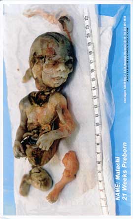
Malachi
Malachi, the literal poster-child for the anti-choice campaign, is a gruesome example of inaccuracy. The back of this 3 x 5 inch card claims that the fetus's life ends in pain. However, judging from the fetus's gray skin, it was aborted via laminaria through an intra-amniotic injection or was done to preserve the mother's life, Andrew Ross. If the procedure was done while the fetus was alive, its skin would be the pinkish color of its left leg
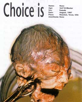
Choice is Abortion
This picture, also distributed on a 3 x 5 inch card and used in numerous anti-abortion campaigns is equally inconsistent. If the forceps securing its head over the jar are standard OR ring forceps, the ring is 1 x 0.5 centimeters, which would mean that the head is too small to be in its third trimester, yet one less developed would not have that much hair, says Dr. Ross. "I strongly suspect that this picture is fake, and the information on the back of the card certainly does not match what's shown," he adds. The second, and most obvious, fallacy lies within the description. It says that the pictured fetus was aborted in 1987 in Texas. However, that was the year that third trimester abortions were banned, rendering the alleged procedure altogether illegal.

Fetal Brain Waves
The National Right to Life Committee released a pamphlet, When Does Life Begin, which states that at six weeks, a fetus has measurable brain waves detected by a an electroencephalogram. But using hard-to-pronounce medical terms to gain credibility does not add substance to this dubious claim. Even at 7-weeks into development, a fetus's brain is the size of a pinhead. Detecting minute brain activity through all the muscle contractions that go on inside a woman's mid-region is a medically unsubstantiated assertion that has not been published in any peer-reviewed journal.
The same pamphlet also cites an article, "What the fetus feels," from the British Medical Journal. This article, lacking any references to studies, supports the organization's claim that the fetus can feel pain. However, in its conclusion, it reads "It is hardly surprising that infants delivered by difficult forceps extraction act as if they have a sever headache." A severe headache?!? How does a newly delivered infant act as if it has a severe headache? This article is simply an example of how anti-choice activists use flawed sources and fallacious information to prove an often medically-unsubstantiated point. For example, Willke, despite outright refutes from The National Cancer Institute, the American Cancer Society, and the American Coalition of Obstetricians and Gynecologists, insists that abortion causes breast cancer.